Rabu, 24 Oktober 2018

VARISELLA (Cacar Air)

No. ICPC II: A72 Chickenpox 
No. ICD X: B01.9Varicella without complication (Varicella NOS) 

Tingkat Kemampuan: 4A 

Masalah Kesehatan 
Varisella atau yang disebut juga dengan bahasa awamnya cacar air merupakan infeksi yang disebabkan oleh virus Varicellazoster yang menyerang kulit dan mukosa. Masa inkubasi 14-21 hari. Penyakit ini dapat menular melalui udara (air-borne) dan kontak langsung. 

Hasil Anamnesis (Subjective) 
Secara umum saat seseorang mengalami penyakit ini, akan mengeluhkan demam, malaise (lemas), dan nyeri kepala. Kemudian disusul timbulnya lesi kulit berupa papul eritem yang dalam waktu beberapa jam berubah menjadi vesikel (plenting-plenting) yang biasanya disertai rasa gatal. Walaupun tidak semua orang yang menderita penyakit ini akan mengalami keluhan serupa seperti yang disebutkan diatas, dikarenakan daya tahan tubuh setiap orang berbeda-beda.

Faktor Risiko 
1. Anak-anak. 
2. Riwayat kontak dengan penderita varisela. 
3. Keadaan imunodefisiensi. 

Hasil Pemeriksaan Fisik dan penunjang sederhana (Objective) 
Pemeriksaan Fisik  
Tanda Patognomonis Erupsi kulit berupa papul eritematosa yang dalam waktu beberapa jam berubah menjadi vesikel. Bentuk vesikel ini khas berupa tetesan embun (tear drops). Vesikel akan menjadi keruh dan kemudian menjadi krusta. Sementara proses ini berlangsung, timbul lagi vesikelvesikel baru yang menimbulkan gambaran polimorfik khas untuk varisela. 


Penyebaran terjadi secara sentrifugal, serta dapat menyerang selaput lendir mata, mulut, dan saluran napas atas. 

Pemeriksaan Penunjang 
Bila diperlukan, pemeriksaan mikroskopis dengan menemukan sel Tzanckyaitu sel datia berinti banyak. 

Penegakan Diagnosis (Assessment) 
Diagnosis Klinis 
Diagnosis ditegakkan berdasarkan anamnesis dan Pemeriksaan Fisik. 

Diagnosis Banding 
1. Variola. 
2. Herpes simpleks disseminata. 
3. Coxsackievirus. 
4. Rickettsialpox. 

Komplikasi 
Pneumonia, ensefalitis, hepatitis, terutama terjadi pada pasien dengan gangguan imun. Varisela pada kehamilan berisiko untuk menyebabkan infeksi intrauterin pada janin, menyebabkan sindrom varisela kongenital. 

Penatalaksanaan komprehensif (Plan) 
Gesekan kulit perlu dihindari agar tidak mengakibatkan pecahnya vesikel. Selain itu, dilakukan pemberian nutrisi TKTP, istirahat dan mencegah kontak dengan orang lain. 

Konseling & Edukasi 
Edukasi bahwa varisella merupakan penyakit yang self-limiting pada anak yang imunokompeten. Komplikasi yang ringan dapat berupa infeksi bakteri sekunder. Oleh karena itu, pasien sebaiknya menjaga kebersihan tubuh. Penderita sebaiknya dikarantina untuk mencegah penularan.  

Kriteria rujukan  
1. Terdapat gangguan imunitas  
2. Mengalami komplikasi yang berat seperti pneumonia, ensefalitis, dan hepatitis. 

Sarana Prasarana 
1. Lup 
2. Laboratorium sederhana untuk pemeriksaan sel Tzanck 

Prognosis 
Prognosis pada pasien dengan imunokompeten adalah bonam, sedangkan pada pasien dengan imunokompromais, prognosis menjadi dubia ad bonam. 


Referensi 
1. Djuanda, A. Hamzah, M. Aisah, S. Buku Ajar Ilmu Penyakit Kulit dan Kelamin, 5th Ed. Balai Penerbit FKUI. Jakarta. 2007. 

2. James, W.D. Berger, T.G. Elston, D.M. Andrew’s  Diseases  of  the  Skin:  Clinical   Dermatology. 10th Ed. Saunders Elsevier. Canada. 2000. 

3. Perhimpunan Dokter Spesialis Kulit dan Kelamin. Pedoman Pelayanan Medik. 2011. 


PANDUAN PRAKTIK KLINIS BAGI DOKTER DI FASILITAS PELAYANAN KESEHATAN PRIMER

Terwujudnya kondisi kesehatan masyarakat yang baik adalah tugas dan tanggung jawab dari negara sebagai bentuk amanah konstitusi yaitu Undang-Undang Dasar Negara Republik Indonesia tahun 1945. Dalam pelaksanaannya negara berkewajiban menjaga mutu pelayanan kesehatan terhadap masyarakat. Mutu pelayanan kesehatan sangat ditentukan oleh fasilitas kesehatan serta tenaga kesehatan yang berkualitas. Untuk mewujudkan tenaga kesehatan yang berkualitas, negara sangat membutuhkan peran organisasi profesi tenaga kesehatan yang memiliki peran menjaga kompetensi anggotanya. 

Bagi tenaga kesehatan dokter, Ikatan Dokter Indonesia yang mendapat amanah untuk menyusun standar profesi bagi seluruh anggotanya, dimulai dari standar etik (Kode Etik Kedokteran Indonesia – KODEKI), standar kompetensi yang merupakan standar minimal yang harus dikuasasi oleh setiap dokter ketika selesai menempuh pendidikan kedokteran, kemudian disusul oleh Standar Pelayanan Kedokteran yang harus dikuasai ketika berada di lokasi pelayanannya, terdiri atas Pedoman Nasional Pelayanan Kedokteran dan Standar Prosedur Operasional.  

Standar Pelayanan Kedokteran merupakan implementasi dalam praktek yang mengacu pada Standar Kompetensi Dokter Indonesia (SKDI). Dalam rangka penjaminan mutu pelayanan, dokter wajib mengikuti kegiatan Pendidikan Pengembangan Keprofesian Berkelanjutan (P2KB) dalam naungan IDI.  

Tingkat kemampuan dokter dalam pengelolaan penyakit di dalam SKDI dikelompokan menjadi 4 tingkatan, yakni : tingkat kemampuan 1, tingkat kemampuan 2, tingkat kemampuan 3A, tingkat kemampuan  3B dan tingkat kemampuan  4B serta tingkat kemampuan 4B. 

Tingkat Kemampuan 1: mengenali dan menjelaskan 
Lulusan dokter mampu mengenali dan menjelaskan gambaran klinik penyakit, dan mengetahui cara yang paling tepat untuk mendapatkan informasi lebih lanjut mengenai penyakit tersebut, selanjutnya menentukan rujukan yang paling tepat bagi pasien. Lulusan dokter juga mampu menindaklanjuti sesudah kembali dari rujukan. 

Tingkat Kemampuan 2: mendiagnosis dan merujuk 
Lulusan dokter mampu membuat diagnosis klinik terhadap penyakit tersebut dan menentukan rujukan yang paling tepat bagi penanganan pasien selanjutnya. Lulusan dokter juga mampu menindaklanjuti sesudah kembali dari rujukan. 

Tingkat Kemampuan 3: mendiagnosis, melakukan penatalaksanaan awal, dan merujuk 

3A. Bukan gawat darurat
Lulusan dokter mampu membuat diagnosis klinik dan memberikan terapi pendahuluan pada keadaan yang bukan gawat darurat. Lulusan dokter mampu menentukan rujukan yang paling tepat bagi penanganan pasien selanjutnya. Lulusan dokter juga mampu menindaklanjuti sesudah kembali dari rujukan. 

3B.Gawat darurat 
Lulusan dokter mampu membuat diagnosis klinik dan memberikan terapi pendahuluan pada keadaan gawat darurat demi menyelamatkan nyawa atau mencegah keparahan dan/atau kecacatan pada pasien. Lulusan dokter mampu menentukan rujukan yang paling tepat bagi penanganan pasien selanjutnya. Lulusan dokter juga mampu menindaklanjuti sesudah kembali dari rujukan. 

Tingkat Kemampuan 4: mendiagnosis, melakukan penatalaksanaan secara mandiri dan tuntas
Lulusan dokter mampu membuat diagnosis klinik dan melakukan penatalaksanaan penyakit tersebut secara mandiri dan tuntas. 
4A. Kompetensi yang dicapai pada saat lulus dokter 
4B. Profisiensi (kemahiran) yang dicapai setelah selesai internsip dan/atau Pendidikan  

Kedokteran Berkelanjutan (PKB) Pada Standar Kompetensi Dokter Indonesia tahun  2012, dari 736 daftar penyakit terdapat 144 penyakit yang harus dikuasai penuh oleh para lulusan karena diharapkan dokter layanan primer dapat mendiagnosis dan melakukan penatalaksanaan secara mandiri dan tuntas. Selain itu terdapat 275 ketrampilan klinik yang juga harus dikuasai oleh lulusan program studi dokter. Selain 144 dari 726 penyakit, juga terdapat 261 penyakit yang harus dikuasai lulusan untuk dapat mendiagnosisnya sebelum kemudian merujuknya, apakah merujuk dalam keadaaan gawat darurat maupun bukan gawat darurat. 

Kondisi saat ini, kasus rujukan ke layanan sekunder untuk kasus-kasus yang seharusnya dapat dituntaskan di layanan primer masih cukup tinggi. Berbagai factor mempengaruhi diantaranya kompetensi dokter, pembiayaan dan sarana prasarana yang belum mendukung. Perlu diketahui pula bahwa sebagian besar penyakit dengan kasus terbanyak di Indonesia berdasarkan Riskesdas 2007 dan 2010 termasuk dalam kriteria 4a.  

Dengan menekankan pada tingkat kemampuan 4, maka dokter layanan primer dapat melaksanakan diagnosis dan menatalaksana penyakit dengan tuntas,. Namun bila pada pasien telah terjadi komplikasi, tingkat keparahan (severity of illness) 3 ke atas, adanya penyakit kronis lain yang sulit dan pasien dengan daya tahan tubuh menurun, yang seluruhnya membutuhkan penanganan lebih lanjut, maka dokter layanan primer secara cepat dan tepat harus membuat pertimbangan dan memutuskan dilakukannya rujukan. 

Melihat kondisi ini, diperlukan adanya panduan bagi dokter pelayanan primer yang merupakan bagian dari standar pelayanan dokter pelayanan primer. Panduan ini selanjutnya menjadi acuan bagi seluruh dokter pelayanan primer dalam menerapkan pelayanan yang bermutu bagi masyarakat. 

Panduan ini diharapkan dapat membantu dokter layanan primer untuk dapat meningkatkan mutu pelayanan sekaligus menurunkan angka rujukan dengan cara: 
  1. Memberi pelayanan sesuai bukti sahih terkini yang cocok dengan kondisi pasien, keluarga dan masyarakatnya 
  2. Menyediakan fasilitas pelayanan sesuai dengan kebutuhan standar pelayanan 
  3. Meningkatkan mawas diri untuk mengembangkan pengetahuan dan ketrampilan professional sesuai dengan kebutuhan pasien dan lingkungan 
  4. Mempertajam kemampuan sebagai gatekeeper pelayanan kedokteran dengan menapis penyakit dalam tahap dini untuk dapat melakukan penatalaksanaan secara cepat dan tepat sebagaimana mestinya layanan primer 


Referensi
Panduan Praktik Klinis Bagi Dokter di Fasilitas Pelayanan Kesehatan Primer Edisi I

Jumat, 14 September 2018

Clinical Care of the HIV-Infected Patient

Primary HIV Infection
Acute retroviral syndrome occurs at the time the infection is acquired in 60% to 80% of HIV infected persons. The illness resembles infectious mononucleosis from infection with Epstein-Barr virus (EBV). Risk factors for transmission of HIV include history of a sexually transmitted disease, especially genital ulcers; unprotected anal or genital intercourse; and multiple sexual partners.

I. Clinical signs and symptoms
A. The period between acquisition of HIV and onset of symptoms is about 14 days, and the characteristic signs and symptoms range from a mild fever and sore throat to a severe mononucleosis-type syndrome with high spiking fevers and a measles-like rash.
B. In those patients with symptomatic seroconversion, the five most common signs and symptoms are fever, fatigue, pharyngitis, weight loss, and myalgias. Characteristic symptoms of acute retroviral syndrome occur in 50% to 90% of patients.

II. Laboratory features
A. Primary HIV infection is diagnosed by a positive plasma HIV RNA obtained on the same day as a negative Western blot assay. When suspicion for acute infection is high, such as in a patient with a report of recent risk behavior in association with symptoms and signs of acute HIV
 nfection, a test for HIV RNA should be performed.
B. Clinical evaluation of possible primary HIV infection often includes a heterophil antibody (Monospot) test to rule out EBV mononucleosis, cytomegalovirus antigen or antibody, acute and convalescent serologic tests for rubella and toxoplasmosis, rapid plasma reagin test, Western blot assay for herpes simplex virus, and serologic tests for hepatitis (including hepatitis C virus RNA polymerase chain reaction).

III. Initial management
A. When the diagnosis of primary HIV has been established, the patient should be examined for syphilis, herpes simplex, venereal warts, gonorrhea, and hepatitis.
B. If the patient was identified as HIV RNA-positive and HIV EIA-negative, a follow-up HIV antibody test should be obtained 2 to 3 weeks after resolution of symptoms to establish that seroconversion has taken place.
C. A baseline CD4+ count should be obtained at the time of diagnosis. In the earliest stages of infection, the CD4+ count can sometimes be below 200 cells/:L. After the first several weeks of infection, a rebound in the CD4+ count to near normal levels may occur.

IV. Treatment of Primary HIV Infection
A. The therapeutic regimen for acute HIV infection should include a combination of two nucleoside reverse transcriptase inhibitors and one potent protease inhibitor. Potential combinations of agents are the same as those used in established infection and include the following regimens:


B. Patient Follow-Up
1.      Testing for plasma HIV RNA levels and CD4+ T cell count and toxicity monitoring should be performed on initiation of therapy, after 4 weeks, and every 3-4 months thereafter.
2.      Antiretroviral agents should be continued indefinitely because viremia has been documented to reappear or increase after discontinuation of therapy.

C. Postexposure prophylaxis
1.      Combination chemotherapy results in fewer transmissions, and use of combination chemotherapy, including a protease inhibitor, is suggested following a significant intravenous exposure.
2.      Postexposure prophylaxis should also be initiated following sexual exposure.
3.      Postexposure prophylaxis treatment regimens
a.                   Zidovudine (ZDV): 300 mg PO bid and
b.                  Lamivudine (3TC, Epivir): 150 mg bid
c.                   Protease Inhibitor: Indinavir (Crixivan) 800 mg q8h or Nelfinavir 750 mg tid (if needed to ensure 2 new antiviral drugs or for very risky exposure).


Selasa, 28 Agustus 2018

Jaundice – Conjugated Hyperbilirubinemia

The presence of jaundice in a child can be a useful indicator of occult pathology. The finding of icterus should set in motion a careful diagnostic search to elucidate the cause. The ultimate goal, to identify precisely the cause of the clinical syndrome, may rest in some cases with the longitudinal caregiver. In all cases, however, the emergency physician at first visit must separate patients whose admission can be temporized from those who require urgent intervention and/or immediate hospitalization.

PATHOPHYSIOLOGY

Unconjugated bilirubin is largely a product of converted heme from senescent red blood cells. Unconjugated bilirubin is transported from extrahepatic reticuloendothelial cells to the liver, bound to albumin. Albumin is detached as the bilirubin gains entry into the hepatocyte. In the liver cell, bilirubin is conjugated with glucuronide by the action of uridine diphosphate glucuronyl transferase. The soluble conjugated diglucuronide then is secreted across the canalicular membrane into the bile. In the intestine, as a result of the activity of bacterial flora, bilirubin is converted to urobilinogen. A portion of urobilinogen is reabsorbed into the portal circulation and is taken up by the liver cells, only to be reexcreted into the bile. A small percentage of urobilinogen escapes into the systemic circulation and is excreted in the urine. The unabsorbed urobilinogen is excreted in the stool as fecal urobilinogen.

In hepatocellular disease, the damaged liver may be unable to excrete the conjugated bilirubin produced in normal amounts. Or, in the absence of hepatic damage, regurgitation into the plasma of conjugated bilirubin may result from functional cholestasis, disruption of the hepatic architecture, or extrahepatic biliary obstruction. In most instances of jaundice primarily related to hepatic disease, the plasma exhibits elevated concentrations of unconjugated and conjugated bilirubin. Overt mechanical obstruction of bile excretion leads to raised plasma levels of conjugated bilirubin, and only as secondary liver damage occurs do unconjugated bilirubin levels rise.

DIFFERENTIAL DIAGNOSIS

Conjugated hyperbilirubinemia is defined by a conjugated bilirubin level higher than 1 mg per dL if the total bilirubin is less than 5 mg per dL or the conjugated bilirubin level represents more than 20% of the total bilirubin if the total bilirubin is higher than 5 mg per dL. Conjugated hyperbilirubinemia, indicating cholestasis, is considered pathological. Cholestatic jaundice may be congenital or acquired. The differential diagnosis includes a variety of structural defects, infections, hepatotoxins, inborn errors of metabolism, and familial syndromes (Table 41.1). Although only a few diseases commonly cause conjugated hyperbilirubinemia (Table 41.2), all are serious. In addition, several less common conditions are important considerations because they are life threatening (Table 41.3).


EVALUATION AND DECISION

It is convenient to divide the approach to patients with conjugated hyperbilirubinemia by age, focusing first on those younger than 8 weeks old and then on those who are older (Fig. 41.1). A majority of children who eventually develop lifethreatening or chronic liver disease initially present in the first 2 months of life. Early physician recognition may lead to successful treatment and a more favorable prognosis.


Infants Younger than 8 Weeks

In the perinatal period, infants develop conjugated hyperbilirubinemia in response to a variety of conditions that may not be encountered in older patients. The increased sensitivity to insult is a result of different patterns of hepatic enzyme activity and liver immaturity with regard to bile formation. Many systemic or hepatic insults may produce perinatal cholestasis (Table 41.1). However, a small number of disorders account for the overwhelming majority of perinatal cholestasis. They include idiopathic neonatal hepatitis, biliary atresia, 1-antitrypsin deficiency, cystic fibrosis, tyrosinemia, galactosemia, choledochal cyst, and perinatal infections. These disorders can be separated by the tempo of the presentation and the appearance of the infant.

The tempo of cholestasis is most abrupt with the infections acquired in utero and during the birthing process. Infected patients are more likely to present shortly after birth. Those who have congenital infections will have a low birth weight. They present with cholestatic jaundice, irritability, jitteriness, and/ or seizures. On examination, microcephaly, hepatomegaly, splenomegaly, and petechiae may be seen with the perinatal TORCHS complex. These include perinatal infections from toxoplasmosis, other infections, rubella, cytomegalovirus (CMV), herpes simplex, and syphilis. Jaundice may be one of the first signs of bacteremia without apparent focus of infection or bacterial sepsis in the first few days of life. Hyperbilirubinemia may occur antecedent to blood cultures becoming positive and may precede findings of anorexia, vomiting, abdominal distension, fever, hepatomegaly, or alterations in respiratory pattern or sensorium. The precise mechanism of jaundice that complicates bacteremia and sepsis is not completely understood. Jaundice may also be an early diagnostic sign of urinary tract infection in the neonatal period. An increase in the conjugated bilirubin fraction may be seen in patients who have afebrile, otherwise asymptomatic urinary tract infection.

The tempo of icterus is subacute with the other common causes of conjugated hyperbilirubinemia. The metabolic and hepatic disorders have variable symptoms at onset. However, the manifestations are far less acute than those seen in the infectious states. Infants with galactosemia, tyrosinemia, and fructose intolerance may appear ill in the emergency department due to metabolic derangement or secondary infection. However, they have had an antecedent history of failure to thrive, developmental delay, and inconstant jaundice. Unexplained fatality in the sibship or unexplained pulmonary, gastrointestinal, neurologic, or psychiatric disturbance in other family members may provoke diagnostic consideration.

The tempo of hepatic and biliary tree disorders is chronic. Those with biliary atresia have intermittent, mild conjugated hyperbilirubinemia during the first 6 to 8 weeks of life. They feed well and thrive. Their stools may be intermittently pigmented early on and become permanently without pigment only after 4 to 6 weeks. They have a benign appearance and with the exception of jaundice and hepatomegaly seem otherwise well. Those patients without a precise anatomic, genetic, or infectious cause of cholestasis are considered to have idiopathic neonatal cholestasis or neonatal hepatitis syndrome. They have onset of their jaundice from 1 to 30 days, with a mean of 7 days. Initially, their stool color is normal, but the stools may become acholic after several weeks. The presence of acholic stools may make it difficult to differentiate between obstructive jaundice causing hepatocellular disease and that caused by obstruction of the biliary tree.

The priorities for the emergency physician are to diagnose medically treatable infections, identify metabolic disorders for which effective therapy is available, and detect extrahepatic obstructive lesions that are amenable to surgical correction. The evaluation begins with cultures of cerebrospinal fluid, blood, urine, and stool. Infants should also have complete blood cell and platelet counts, coagulation studies, hepatic enzymes (aspartate aminotransferase, alanine aminotransferase, and gamma-glutamyl transpeptidase), ammonia, albumin, total protein, alkaline phosphatase, electrolytes, blood urea nitrogen, creatinine, and blood sugar tests. Urine should be obtained for urinalysis and tested for reducing substances. A right upper quadrant ultrasound should be performed in order to identify anatomic abnormalities such as a choledochal cyst. If cystic fibrosis is suspected, schedule the patient for a sweat chloride iontophoresis or testing for common genetic variants in the cystic fibrosis transmembrane conductance regulator gene. Additional studies of blood and urine that they may find useful include 1-antitrypsin, TORCHS and hepatitis B virus serology, serum amino acids, thyroid function tests, red blood cell galactose 1-phosphate uridyltransferase activity, and urine examination for cytomegalovirus.

Inpatient observation is appropriate in this age group because the diagnosis can rarely be established in the emergency department. Empiric therapy for sepsis or urinary infection is often warranted, pending culture results.

Children Older than 8 Weeks

In the evaluation of conjugated hyperbilirubinemia beyond infancy, it is necessary to know if there has been exposure to contagion or a potential for sexual or vertical transmission of infections such as hepatitis or human immunodeficiency virus. Other risk factors for hepatitis (e.g., needle sticks, hemodialysis, transplant, transfusion of blood products, or factor use) need to be evaluated. The physician should pursue possible exposure to industrial toxins or foods previously implicated in hepatic injury (e.g., carbon tetrachloride, yellow phosphorus, tannic acid, alcohol, mushrooms of the Amanita species). The emergency physician must inquire about use of acetaminophen, salicylates, nonsteroidal anti-inflammatory drugs, iron salts, erythromycin estolate, ceftriaxone, rifampin, nitrofurantoin, oxacillin, tetracycline, trimethoprim-sulfamethoxazole, ketoconazole, diphenylhydantoin, isoniazid, and chlorpromazine. The presence of prior episodes of jaundice, acholic stools, and/or abdominal pain may suggest an underlying disorder, predisposing the patient to obstruction of the biliary tree. Other historical points include the presence of fever, arthralgia, arthritis, conjunctivitis, rash, pruritus, vomiting, diarrhea, weight loss, color of the urine, abnormal bruising or spontaneous bleeding, and changes in mental status. An examination that focuses on ongoing physical signs of liver disease may result in greater accuracy in clinical evaluation of the older jaundiced patient. These signs include skin changes (spider angiomata, excoriations, palmar erythema) and peripheral edema. The abdominal examination should include observations of the venous pattern, presence of ascites, mass, or peritoneal irritation. There should be an estimation of liver size, contour, and tenderness, as well as an estimate of spleen size. The clinician should exclude cardiovascular dysfunctions such as hypoxemia, systemic venous congestion, and low cardiac output. Observations should be made of mental status and neuromuscular changes. Patients with cystic fibrosis, 1-antitrypsin deficiency, Wilson’s disease, or inflammatory bowel disease tend to have symptoms that remit and relax. However, slow progression is the rule. Patients with 1-antitrypsin deficiency may have onset of respiratory or hepatic complaints at any age. Similarly, infants who have failure to thrive from cystic fibrosis may develop obstruction at any age in the extrahepatic or intrahepatic ducts and, transiently or persistently, may exhibit jaundice. Patients with ulcerative colitis and Crohn’s disease may become symptomatic intermittently with episodes of cholestasis. The degree of hepatic derangement and expression of neurologic abnormality is variable with Wilson’s disease. Before the diagnosis is entertained, patients typically exhibit dysarthria, tremors, rigidity, or psychic disturbances. Rarely, younger patients without prodromal events have acute jaundice and hepatomegaly and progress to hepatic failure. Biliary calculi and acute inflammation of the gallbladder are uncommon causes of conjugated hyperbilirubinemia in the pediatric population. However, a subset of patients is predisposed to these complications. Cholelithiasis may complicate any of the hemolytic anemias, particularly in patients with sickle hemoglobinopathies. These patients have increased incidence of both liver and gallbladder disease. Liver or gallbladder dysfunction accounts for jaundice when more than 10% of an elevated bilirubin in a patient with sickle cell disease is conjugated. Cholecystitis may accompany a variety of acute focal infections, such as pneumonia or peritonitis, and may occur in the course of bacterial sepsis. In this event, shock and hyperpyrexia may divert the clinician from the deranged biliary system. In less severe cases, fever, nausea, vomiting, abdominal distension, and right upper quadrant pain are prominent features of cholecystitis. Right upper quadrant abdominal mass, pain, and jaundice constitute the classic triad in the diagnosis of choledochal cyst. The clinical recognition may be delayed until there is a complication, such as cholangitis. An acute, painful right upper quadrant mass associated with jaundice may also occur in the course of acute hydrops of the gallbladder from Kawasaki disease or systemic streptococcal infection. In the previously healthy child, the most common cause of conjugated hyperbilirubinemia is acute hepatitis . The illness may be abrupt in onset, with fever, urticaria, and arthralgia as primary manifestations. More often, the illness is insidious. Viral hepatitis is characterized by low-grade fever and gastrointestinal complaints such as anorexia, malaise, nausea, vomiting, and abdominal pain before the jaundice. Liver enlargement with hepatitis (A, B, C, and non-A, non-B, non-C), varicella, herpesvirus, Coxsackievirus, echovirus, Epstein-Barr virus, and adenovirus infection is inconstant. Hepatic tenderness is a more reliable finding. Rarely, ascites can accompany hepatitis virus infection. Splenomegaly is the rule with Epstein-Barr virus but is unusual with the other agents. On occasion, hepatitis may be associated with a distinctive erythematous papular eruption localized to the limbs (Gianotti-Crosti syndrome). Toxic hepatitis, unlike viral hepatitis, does not have a prolonged prodrome. Acute nausea, vomiting, and malaise are followed in 1 to 2 days by alterations in mental status and deterioration of liver function. Most patients with toxic hepatitis will have an identifiable exogenous precipitant. Children with fulminant hepatic failure typically experience anorexia, nausea, vomiting, malaise, and fatigue—all symptoms indistinguishable from those expected with viral hepatitis. The patient’s jaundice becomes more profound and vomiting becomes protracted. Hyperexcitability, mania, and subtle psychomotor abnormalities may be seen. Coagulopathy, ascites, and sudden decrease in liver size are often the prelude to the development of frank neuromuscular signs. The objectives of the emergency physician are to render supportive care to those icteric patients with infectious and metabolic derangements and to identify those cases in which jaundice is caused by mechanical obstruction or hepatic failure. The impression based on a targeted history, physical examination, and clinical algorithms can be bolstered with the following laboratory examinations: complete blood cell count, platelet count, coagulation profile, prothrombin time, total and direct bilirubin, hepatic enzymes, alkaline phosphatase, electrolytes, blood urea nitrogen, and creatinine. Urinalysis, culture, and toxicologic screen should be considered. Chest and abdominal radiographs are indicated when there are pulmonary parenchymal complaints or significant abdominal findings. Other laboratory tests that are often available immediately and that may provide useful information in specific circumstances are serum ammonia, albumin, total protein, lipid profile, pH, and carbon dioxide. If available, abdominal sonography or computed tomography may be helpful occasionally. In no circumstance will results of several important blood and urine tests be of immediate use. Such studies, which are appropriate, include antinuclear antibody, fetoprotein, and serum for bile acids, ceruloplasmin, protein electrophoretic pattern, polymerase chain reaction assays or serologic evidence of recent infection (e.g., Epstein-Barr virus, mycoplasmal or hepatitis profiles). Urinary analysis includes assessment of organic acids and copper. These investigations may be helpful, however, to the longitudinal caregiver who must maintain a vigilant watch over the jaundiced patient. Children older than 8 weeks with conjugated hyperbilirubinemia should be admitted to the hospital at the time of their presentation in all cases in which life-threatening conditions may exist (Table 41.3). Inpatient treatment is also suggested when intravenous fluids are necessary to treat symptomatic hypoglycemia or electrolyte imbalance and when operative intervention may prove necessary. Icteric patients who have been diagnosed previously with confidence and who have exacerbation of their symptoms may require admission to reappraise their status. The physician may also be influenced to admit the patient when social factors or geographic barriers inhibit consistent observations. Admission is also indicated for patients who require further diagnostic intervention, such as hepatobiliary scintigraphy, endoscopic retrograde cholangiopancreatography, or liver biopsy to arrive at a definitive diagnosis.

Suggested Readings

Crain EF, Gershel JC. Urinary tract infections in febrile infants younger than 8 weeks of age. Pediatrics 1990;86:363–367.

Emerick KM, Whitington PF. Neonatal liver disease. Pediatr Ann 2006;35: 280–286.

Garcia FJ, Nager AL. Jaundice as an early diagnostic sign of urinary tract infection in infancy. Pediatrics 2002;109:846–851.

Jacquemin E, Lykavieris P, Chaoui N, et al. Transient neonatal cholestasis: origin and outcome. J Pediatr 1998;133:563–567.

Moyer V, Freese DK, Whitington PF, et al. Guideline for the evaluation of cholestatic jaundice in infants: recommendations of the North American Society for Pediatric Gastroenterology, Hepatology and Nutrition. J Pediatr Gastroenterol Nutr 2004;39:115–128.


Suchy FJ. Approach to the infant with cholestasis. In: Suchy FJ, Balistreri WF, Sokol RJ, eds. Liver disease in children, 3rd ed. New York, NY: Cambridge University Press, 2007:187–194.

Constipation In Pediatric

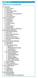
Constipation is an important problem in the pediatric emergency department for many reasons. It is one of the most common pediatric complaints, accounting for 3% of primary care visits. There are many causes for constipation (Table 13.1), some rare and some very common (Table 13.2). Occasionally, the presentation of constipation is atypical, with chief complaints that superficially seem unrelated to the gastrointestinal tract (Table 13.3). Although relatively rare, some causes of constipation are potentially life threatening and need to be recognized promptly by the emergency physician (Table 13.4). In addition, constipation may produce symptoms that mimic other serious illnesses such as appendicitis.


DEFINITION

Although constipation most commonly is defined as decreased stool frequency, there is not one simple definition. The stooling pattern of children changes based on age, diet, and other factors. Average stooling frequency in infants is approximately 4 stools per day during the first week of life, decreasing to 1.7 stools per day by 2 years of age, and approaching the adult frequency of 1.2 stools per day by 4 years of age. Nevertheless, normal infants can range from 7 stools per day to 1 stool per week. Older children can defecate every 2 to 3 days and be normal.

It is easier to define constipation as a problem with defecation. This may encompass infrequent stooling, passage of large and/or hard stools associated with pain, incomplete evacuation of rectal contents, involuntary soiling (encopresis), or inability to pass stool at all.

PHYSIOLOGY

The passage of food from mouth to anus is a complex process. The intestine relies on input from intrinsic nerves, extrinsic nerves, and hormones to function properly. Normal defecation involves voluntary and involuntary components. Disruption of any of these can result in constipation. The colon is specialized to transport fecal material and balance water and electrolytes contained in the feces. When all is functioning well, the fecal bolus arrives in the rectum formed but soft enough for easy passage through the anus. Normal defecation requires the coordination of the autonomic and somatic nervous systems and normal anatomy of the anorectal region. The internal anal sphincter is a smooth muscle, which is innervated by the autonomic nervous system. It is tonically contracted at baseline. It relaxes in response to the arrival of a fecal bolus in the rectum, allowing stool to descend to the portion of the anus innervated by somatic nerves. At this point, the external anal sphincter, striated muscle under voluntary control, tightens until the appropriate time for fecal passage. Before defecation, squatting straightens the angle between the rectum and the anal canal, allowing easier passage. Voluntary relaxation of the external anal sphincter allows passage of the feces, and increasing intraabdominal pressure via Valsalva aids the process.

EVALUATION AND DECISION

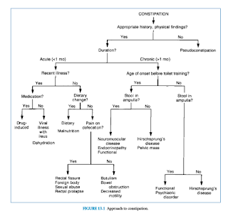
The evaluation of the child presumed to have constipation should begin with a thorough history and physical examination. Special attention should be paid to the age of the patient, duration of symptoms, timing of first meconium passage after birth, changes in frequency and consistency of stool, stool incontinence, pain with defecation, rectal bleeding, presence of abdominal distention and/or palpable feces, and a rectal exam to assess anal position, sphincter tone, widening of the rectal vault, and presence of hard stool. A complaint of constipation is not sufficient for diagnosis. A decrease in stool frequency or the appearance of straining is often interpreted as constipation. The physician should be aware of the grunting baby syndrome, or infant dyschezia, in which an infant grunts, turns red, strains, and may cry while passing a soft stool. This is the result of poor coordination between Valsalva and relaxation of the voluntary sphincter muscles. Examination reveals the absence of palpable stool in the rectum or abdomen. Complaints of constipation not supported by history or physical examination are called pseudoconstipation (Fig. 13.1).
Acute Constipation

Constipation is not a disease; it is a symptom of a problem. Constipation is acute when it has occurred for less than 1 month’s duration. The patient’s age and the duration of the constipation are important when determining the cause and significance of the problem.

The infant younger than 1 year of age with true constipation is particularly concerning. Potential causes include serious diseases such as dehydration, malnutrition, and infant botulism. A recent viral illness accompanied by dehydration from excessive water loss through vomiting, diarrhea, fever, and increased respiratory rate can precipitate acute constipation in an infant. Adynamic ileus or decreased intake after gastroenteritis may cause slower transit time through the colon, which can also lead to hard stools. Anal fissures and/or diaper rash after a bout of diarrhea may precipitate painful defecation, resulting in stool retention. In this case, the infant may assume a retentive posture consisting of extension of the body with contraction of the gluteal and anal muscles.

Excessive intake of cow’s milk, inadequate fluid intake, and malnutrition should all be uncovered by a complete dietary history. Recent courses of medication cannot be overlooked because many can cause constipation (Table 13.5). Ingestion of lead is also a potential and serious reason for constipation.


Infantile botulism commonly presents with acute constipation, weak cry, poor feeding, and decreasing muscle tone. Acute constipation can also be a symptom of a bowel obstruction, but is normally a less prominent feature than other symptoms.

Acute constipation in the child older than 1 year of age occurs for many of the same reasons as in the infant. History may reveal recent viral illness or use of medication, as well as the presence of underlying illness, such as neuromuscular disease. Physical examination suffices to rule out anal malformations and other physical problems that could result in trouble defecating.

Chronic Constipation

Constipation of more than 1 month’s duration in an infant, although probably a functional problem, is especially concerning and should prompt consideration of an underlying illness. Spinal muscular atrophy, amyotonia, congenital absence of abdominal muscles, dystonic states, and spinal dysraphism, which cause problems with defecation, can be readily diagnosed with history and physical examination.

Anorectal anomalies occur in approximately 1 in 2,500 live births. Anal stenosis causes the passage of ribbonlike stools with intense effort. Diagnosis is made by anal examination, which demonstrates a tight, constricted canal. The condition is treated by repeated anal dilations, sometimes over several months. The anus can be covered by a flap of skin, leaving only a portion open for passage of stool. This “covered anus” may require anoplasty with dilation. Anterior displacement of the anus is believed to cause constipation by creating a pouch at the posterior portion of the distal rectum that catches the stool and allows only overflow to be expelled after great straining. The treatment may be medical or surgical.

Hirschsprung’s disease, or congenital intestinal aganglionosis, is rare but must be considered in the constipated infant because it has the potential to cause life-threatening complications. The incidence is 1 in 5,000 live births, with a male:female predominance of 4:1. As a result of failure of migration of ganglion cell precursors along the gastrointestinal tract, there is the absence of ganglion cells in the submucosal and myenteric plexuses of the affected segment. The absence of ganglion cells leaves the affected segment tonically contracted, blocking passage of stool. The segment proximal to the blockage dilates as the buildup of stool progresses. In most cases, the child never feels the urge to defecate because the blockage is proximal to the internal sphincter and anal canal.

In Hirschsprung’s disease, abdominal examination often yields a suprapubic mass of stool that may extend throughout the abdomen. Rectal examination reveals a constricted anal canal with the absence of stool in the rectal vault, commonly followed by expulsion of stool when the finger is removed. The combination of palpable abdominal feces and an empty rectal vault is abnormal and must be further investigated.

Megacolon in Hirschsprung’s disease can lead to enterocolitis characterized by abdominal distension; explosive stools, which are sometimes bloody; and fever progressing to sepsis and hypovolemic shock. Enterocolitis represents a major cause of mortality in this condition.

Of infants with Hirschsprung’s disease, 80% are diagnosed within the first year of life. A history of late passage of meconium is often found (Table 13.6). However, if the involved segment is relatively short, the diagnosis may be delayed. If suspected, diagnosis is supported by unprepped barium enema, which typically demonstrates narrow bowel rapidly expanding to a dilated area. This transition zone represents the location where the aganglionic, tonically contracted bowel meets the dilated, innervated bowel. In disease where only a short segment of bowel is involved, barium enema may miss the transition zone and anal manometry aids in diagnosis. Confirmation is achieved by demonstration of aganglionosis on biopsy.


Hypothyroidism in the infant may present with constipation. Water-losing disorders such as diabetes insipidus and renal tubular acidosis may also contribute to this condition. Cystic fibrosis can present with constipation alone; when there is a history of delayed passage of meconium and Hirschsprung’s disease has been ruled out, evaluation by a sweat test is indicated.

Chronic constipation in the older child is overwhelmingly likely to be functional constipation. Typically, a cycle of stoolwithholding starts when the child disregards the signal to defecate and strikes a retentive posture—rising on the toes and stiffening the legs and buttocks. This maneuver forces the stool out of the anal canal and back into the rectum, which subjects the fecal bolus to further absorption of water. The longer the stool sits, the more likely defecation is to be painful and traumatic. This reinforces stool-withholding behavior, creating larger and harder stool in the rectum.

Over time, in functional constipation, the rectum dilates and sensation diminishes. Eventually, the child loses the urge to defecate altogether. Watery stool from higher in the gastrointestinal tract can leak around the large fecal mass, causing involuntary soiling, or encopresis. This may be misconstrued as diarrhea or as regression in the toilet-trained child. Many parents consult a physician at this point. Other reasons parents seek medical attention for their children are abdominal pain, anorexia, vomiting, and irritability.

Peak times for constipation to develop are when routines change. Toilet training represents a major alteration in the toddler’s routine. It is also a time when the child and caregiver battle for control. Another problematic time is after starting school, when a child may be uncomfortable using an unfamiliar bathroom or unable to adapt to a lack of privacy. Involvement with friends or games may distract a child from the signal to defecate. Painful defecation from streptococcal perianal disease or sexual abuse must be remembered as potential precipitants of stool withholding. In addition, functional constipation can be associated with dysfunctional urinary voiding and urinary tract infections.

A history supportive of functional constipation includes retentive posturing, infrequent passage of very large stools, and involuntary soiling during the peak ages. Physical examination typically reveals palpable stool in the abdomen. The back should be inspected for skin changes over the sacral area, which would suggest spinal dysraphism. Normal deep tendon reflexes and strength in the lower extremities in conjunction with a normal anal-wink reflex virtually excludes neurologic impairment. The anus should be normal in placement and appearance. Rectal examination typically yields a dilated vault filled with stool. Abdominal flat-plate x-ray can be helpful but is not necessary (Fig. 13.2). Failure to thrive is not associated with functional constipation and, if present, should prompt further investigation.

Although functional constipation encompasses most cases of chronic constipation in the child older than 1 year of age, the less common causes must always be considered.

As in the infant, endocrine abnormalities and other disorders can cause and present as constipation. Hypothyroidism is often associated with constipation, as well as with sluggishness, somnolence, hypothermia, weight gain, and peripheral edema. Diabetes mellitus produces increased urinary water loss and, in the long term, intestinal dysmotility, which can lead to constipation. Hyperparathyroidism and hypervitaminosis D, which lead to increased serum calcium, cause constipation through decreased peristalsis. Celiac disease is also recognized as a cause of chronic constipation.

Rarely, an abdominal or pelvic mass may present with chronic constipation. Careful abdominal examination will demonstrate the mass. Rectal masses may present similarly. Follow-up again is emphasized because a mass that does not resolve after clearance of impaction needs further evaluation. Hydrometrocolpos can present with constipation and urinary frequency; therefore, a genital examination is indicated in girls to document a perforated hymen. One must also remember that intrauterine pregnancy is a common cause of pelvic mass and constipation in adolescent girls.

Children with neuromuscular disorders often develop chronic constipation. Myasthenia gravis, the muscular dystrophies, and other dystonic states can predispose children to constipation through a number of mechanisms. A detailed history and physical examination should recognize most neuromuscular problems, allowing symptomatic treatment to be provided.

Psychiatric problems must not be forgotten in the evaluation of constipation. Depression can be associated with constipation secondary to decreased intake, irregular diet, and decreased activity. Many psychotropic drugs can cause constipation. Anorexia nervosa may present with constipation because of decreased intake or metabolic abnormalities, and laxative abuse can cause paradoxical constipation.

TREATMENT

Simple acute constipation in an infant should be treated initially with dietary changes (Table 13.7). Decreasing consumption of cow’s milk, possible formula change, and increasing fluid intake when appropriate may be enough to alleviate the symptoms. In addition, supplementing the diet with sorbitol as found in prune, pear, white grape, and apple juice can be helpful to soften the stool and improve stool passage. If dietary measures are not sufficient, lactulose or barley malt soup extract (Maltsupex®) may be useful as osmotic agents. Historically, Karo® corn syrup had been used as an osmotic agent, but its use has fallen out of favor after concerns that the syrup may contain spores of Clostridium botulinum. Stool lubricants such as mineral oil should not be used in children younger than 3 years of age and should also be avoided in some older children when aspiration is a risk. Polyethylene glycol solutions such as MiraLax® have gained increased use in the outpatient setting (see discussion below). When perianal irritation or anal fissures are present, local perianal care may decrease the risk of painful defecation, which, in turn, may decrease stool-retentive behavior. Follow-up is the most important aspect of treating simple constipation.

Therapy for acute functional constipation in the child older than 1 year of age should be the same as that for the infant, with dietary changes and stool softeners as mainstays; however, attention should also be paid to psychological factors such as recent stress that may be complicating the situation.

Treatment for chronic constipation in the infant younger than 1 year of age should include ongoing dietary measures including several daily servings of pureed fruits and vegetables, sorbitol-containing juices, and possible formula change. If dietary measures alone are insufficient to control symptoms, a daily stool softener such as lactulose can be used to help maintain soft stool passage and a glycerin suppository can be used on occasion to disimpact the rectum, although this should not be used regularly. Although safety data are still emerging, polyethylene glycol (PEG) 3350 (MiraLax®, GlycoLax®) may also be a safe and effective treatment for chronic constipation in infants. Loening-Baucke and colleagues studied 20 children and Michail and colleagues studied 12 children younger than 1 year of age, who were safely and successfully treated with PEG 3350 used for several months or more. Although more safety data is needed to make specific recommendations, this will likely become one of the therapeutic options for this age range.

Treatment (Table 13.7) for chronic functional constipation in the child older than 1 year of age begins with disimpaction and evacuation of the stool remaining in the colon. This is accomplished with either oral or rectal therapy or a combination of the two. A study by Youssef et al. demonstrated that in fecally impacted children whose palpable stool mass did not extend above the level of the umbilicus, PEG 3350  at a dose of 1 to 1.5 g per kg per day (up to a maximum of 100 g per day) given for 3 days was an effective method of disimpaction and evacuation. Other oral options include lactulose, sorbitol, senna, bisacodyl, PEG electrolyte solution, magnesium hydroxide, and magnesium citrate. Rectal disimpaction can be accomplished with hypertonic phosphate (Fleet®) enemas or bisocodyl suppositories. A mineral oil enema administered the night before the first phosphate enema may soften existing stool, allowing less painful passage. Phosphate enemas are typically dosed at one adult-sized enema (133 mL) for patients 3 years and older, and one pediatric-sized enema (66 mL) for those 1 to 3 years of age. Phosphate enemas should not be used in children younger than 1 year. The enema may be repeated, spaced 24 hours apart, with a maximum of three total doses. Subsequent doses should only be given if evacuation of the previous dose has occurred. Phosphate enemas should be used with caution in patients with dehydration, prolonged enema retention, or renal impairment because such use has rarely been associated with severe hyperphosphatemia, hypocalcemia, and tetany, and consequent life-threatening complications. Tap water and soapsuds enemas should be avoided because of the possibility of water intoxication. Enemas will disimpact but oral agents are often needed in addition to produce full bowel evacuation. If there is no response after 2 days, more aggressive disimpaction under physician supervision is indicated.  Oral phospho-soda preparations should never be used in children and have been removed from the market secondary to serious electrolyte abnormalities.

The long-term maintenance phase of therapy, which is equally as important as the disimpaction and evacuation phase, involves nonstimulant osmotic laxatives, lubricants, fluids, fiber, and behavioral therapy. Laxatives include hyperosmolar agents such as lactulose and PEG 3350. Lubricants such as mineral oil and Kondremul® are helpful to lubricate the intestine for easier passage of stool. These should only be used in children older than 3 years of age and those without a high risk for aspiration. Some have advocated the use of fatsoluble vitamin supplementation when mineral oil products are used, but there is little evidence to suggest this is truly necessary. Increasing fluid and fiber intake is also critical to longterm success in treating constipation. Table 13.8 outlines the recommended daily fiber intake for different ages. Fiber should be increased gradually toward the goal to minimize side effects of flatulence. Regular toileting should be encouraged with positive reinforcement in the school-age child. Toilet training should be discontinued in the training toddler until retentive behaviors have improved. Education of patients and parents about the pathophysiology of constipation, the etiology of encopresis when present, and the expectations of therapy are vital. Close follow-up is a mainstay of treatment. Successful therapy may take months to years to complete.

Approach to the Patient with Severe Chronic Constipation

Disimpaction and evacuation of stool in the patient with severe chronic constipation or one who has failed simple therapy presents a challenge, particularly in the emergency department setting. A series of phosphate enemas may not be sufficient to disimpact a larger stool mass. Use of PEG with electrolytes solution (GoLYTELY®) as a lavage either orally or via nasogastric tube at a dose of 10 to 25 mL per kg per hour up to 1000 mL per hour until stool is clear may be helpful to treat more severe impactions. This method should be done in the hospital under supervision of a physician with close monitoring of the patient’s volume and cardiovascular status and electrolytes. Risks may be higher in patients with complex medical conditions such as cardiac disease. Gastrograffin or N-acetylcysteine enemas may be an additional method of disimpaction, especially in the case of distal intestinal obstructive syndrome as occurs in patients with cystic fibrosis. In cases of very severe fecal impaction, surgical disimpaction may be necessary. The use of milk and molasses enemas in children is falling out of favor in many institutions as a result of safety concerns following several case reports of serious adverse events, including one death, after administration. The other components of constipation therapy apply as already outlined previously and in Table 13.7.


Suggested Readings

Benninga MA, Voskuijl WP, Taminiau JAJM. Childhood constipation: Is there new light in the tunnel? J Pediatr Gastroenterol Nutr 2004;39:448–464.

Constipation Guideline Committee of the North American Society for Pediatric Gastroenterology, Hepatology and Nutrition. Evaluation and treatment of constipation in infants and children: recommendations of the North American Society for Pediatric Gastroenterology, Hepatology and Nutrition. J Pediatr Gastroenterol Nutr 2006;43:e1–e13.

Loening-Baucke V. Prevalence, symptoms and outcome of constipation in infants and toddlers. J Pediatr 2005;146:359–363.

Loening-Baucke V, Krishna R, Pashankar DS. Polyethylene glycol 3350 without electrolytes for the treatment of functional constipation in infants and toddlers. J Pediatr Gastroenterol Nutr 2004;39:536–539.

Michail S, Gendy E, Preud’Homme D, et al. Polyethylene glycol for constipation in children younger than eighteen months old. J Pediatr Gastroenterol Nutr 2004;39:197–199.

Nurko S, Youssef NN, Sabri M, et al. PEG3350 in the treatment of childhood constipation: a multicenter, double-blinded, placebo-controlled trial. J Pediatr 2008;153:254–261.

Pashankar D, Loening-Baucke V, Bishop W. Safety of polyethylene glycol 3350 for the treatment of chronic constipation in children. Arch Pediatr Adolesc Med 2003;157:661–664.

Walker M, Warner BW, Brilli RJ, Jacobs BR. Cardiopulmonary compromise associated with milk and molasses enema use in children. J Pediatr Gastroenterol Nutr 2003;36:144–148.


Youssef N, Peters JM, Henderson W, et al. Dose response of PEG 3350 for the treatment of childhood fecal impaction. J Pediatr 2002;141(3):410–414.